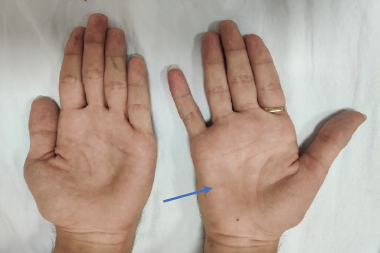
Diabetes mellitus (DM) is a leading cause of peripheral neuropathy.1–3 A distal symmetric sensorimotor polyneuropathy (DSP), is the most common manifestation,4 and is considered to be mainly due to axonal degeneration and progressive loss of nerve fibers.5–7 However, focal and multifocal peripheral nerve lesions, comprising cranial, thoracoabdominal, and limb nerve lesions, including proximal lumbosacral radiculoplexus neuropathies, can also occur.8 In contrast to the common DSP phenotype, chronic inflammatory demyelinating polyneuropathy (CIDP) is characterized by motor greater than sensory, proximal, and distal peripheral neuropathy, with a slowly progressive or relapsing course.9 It is also associated with impaired sensation, absent or diminished tendon reflexes, and elevated cerebrospinal fluid (CSF) protein level, as well as demyelinating features observed on nerve conduction studies.10 However, different variants exist, including a primary sensory ataxic form.11,12 Therapy should be initiated early in the course of the disease to prevent ongoing demyelination and secondary axonal loss leading to permanent disability.10 Although there are similar treatment response rates between patients with diabetes (CIDP+DM) and without diabetes (CIDP-DM),13–16 CIDP+DM patients are less likely to receive immune therapies.13,15 This is possibly due to the greater challenge of diagnosing CIDP in DM patients who likely also have DSP. The difficulty in the diagnosis might be attributable to CIDP clinical heterogeneity, multifocality, predilection for proximal nerve segments, and the limitations of electrophysiologic and pathologic investigations in distinguishing between primary demyelinating and axonal processes.17 Although there are abundant research criteria for the diagnosis CIDP,18 and these may be appropriate in the research setting, such rigorous electrophysiologic criteria lack sensitivity for the diagnosis, and may miss clinical cases of CIDP.9 Currently, there are no widely accepted practical clinical criteria on which to base treatment.17
Epidemiology
DM is pandemic, with a prevalence of 8.3 % as per the International Diabetes Federation’s Diabetes Atlas 2012; however, it is estimated that up to half of all cases have not been diagnosed.19 In those greater than 65 years of age, the prevalence is 27 % and expected to climb greatly by 2050 if current trends continue.20 About 20 % of patients with diabetes duration less than 5 years have clinical peripheral neuropathy.21 In type 1 patients, the prevalence of DSP is estimated to be 28 %.22 This rate increases to at least 50 % among patients who have had diabetes for 25 years,5,21 resulting in DM becoming the leading cause of peripheral neuropathy.1–3 By contrast, CIDP is relatively uncommon, although it is considered to be the most common chronic autoimmune neuropathy.23 Previously published data on its prevalence in the general population varied greatly, with estimates ranging from one to 8.9 cases per 100,000;9,24,25 however, the actual prevalence might be greatly underestimated.17 The question of whether there is higher prevalence of CIDP in DM is controversial.
In a prospective study, Sharma et al. found a significantly higher occurrence of demyelinating neuropathy meeting the electrophysiologic criteria for CIDP in types 1 and 2 DM patients (32/189 DM patients, 16.9 %), than in nondiabetic patients (17/938 patients, 1.8 %), with a calculated odds of occurrence 11 times higher among diabetic than nondiabetic patients. The odds for DM in CIDP patients was also found to be 20 times higher than in myasthenia gravis (MG) and amyotrophic lateral sclerosis (ALS) patients.26 Similarly, in a retrospective review of 87 CIDP patients, Rotta et al. found a high percentage of patients with DM of 26 %.15 By contrast, in a cohort of 155 patients with CIDP, Chiò et al. found only 14 (9.0 %) DM patients, including 12 type 2 and two type 1 patients (close to the predicted number of 13.03), concluding that there is no pathogenetic correlation between the two.27 Similarly, although in a smaller study, Laughlin et al. found that only one of 23 CIDP patients (4 %) had DM, whereas 14 of 115 age- and sex-matched controls (12 %) had DM, concluding that DM is not a major risk factor in the development of CIDP. The perceived association of DM with CIDP was suggested to be due to a chance association or misidentification of other forms of diabetic neuropathy.9 Any association between CIDP and DM is unclear at this time and part of this difficulty might arise from the numerous criteria available to make the diagnosis of CIDP.
Pathogenesis
Progressive loss of nerve fibers is the hallmark of DSP4 as reflected in the electrophysiology.28 Nonetheless, Dunnigan et al. showed that DSP can be classified into different pathophysiologic types of axonal, conduction slowing, or combined DSP with different clinical characteristics, supporting the hypothesis that pathophysiologic differences may exist within the spectrum of DSP (see Table 1). Evidence of conduction slowing (likely demyelination) was found to be associated with worse glycemic control in patients with type 1 diabetes, demonstrating that metabolic factors can determine different pathophysiologic behaviors.29 Microangiopathy is observed commonly, and is occasionally associated with potentially reversible metabolic, immunologic, or ischemic injury.30 It has been suggested that diabetic nerve damage can expose peripheral nerve antigens to the immune system, and consequently DSP might be a predisposing event to immune-mediated neuropathies.7 Given that recent work suggests a demyelinating component in type 1 DM patients with poor glycemic control, an adverse inflammatory process might be caused by the metabolic state.29 In CIDP there is a progressive loss of immunologic tolerance to peripheral nerve components such as myelin, Schwann cell, the axon, and motor or ganglionic neurons. Activated macrophages, T cells, and auto-antibodies induce an immune attack against peripheral nerve antigens. Complement-fixing immunoglobulin deposits are localized to the myelin sheath surrounding axons.10,23 Activated tissue macrophages comprise the final process of demyelination by invading the lamellae causing focal damage to the myelin sheath.31 Concomitant axonal loss secondary to primary demyelination is common.32
Clinical Manifestations
Diabetic neuropathy has several distinct forms. The most common form is a chronic, predominantly sensory DSP, which is often painful, especially in severe forms,33 but rarely produces major weakness on physical examination.7 By contrast, CIDP is characterized by motor greater than sensory, proximal, and distal peripheral neuropathy, and is often painless.9 CIDP, by definition, progresses for more than 2 months, and is associated with impaired sensation, and absent or diminished tendon reflexes.10 The classic presentation does not address well-recognized variants, such as those with predominantly distal involvement, cranial nerve palsies, exclusively sensory polyneuropathy, markedly asymmetric disease, and even associated central nervous system (CNS) demyelination.15 CIDP+DM patients have a longer delay from onset to diagnosis, but do not differ in the mean age of onset, gender distribution, and the type of clinical course.15,27 However, worse clinical manifestations can be expected in CIDP+DM patients compared with CIDP-DM patients, as they reflect the consequence of two different pathogenic processes affecting the nerves.13 Gorson et al. found that complaints of imbalance were more frequent in CIDP+DM patients.14 Similarly, Dunnigan et al. found that CIDP+DM subjects had more severe neuropathy based on more proximal weakness, higher Toronto Clinical Neuropathy Score (TCNS), more gait abnormality, and higher lower limb vibration potential thresholds (VPT).13 In DSP patients with probable demyelination related to poor glycemic control compared with CIDP+DM patients (see Table 2), the DSP patients had less severe neuropathy, a longer duration of diabetes, and worse glycemic control suggesting differing etiologies for these entities, despite similarities in the electrophysiologic pattern of demyelination.34
Electrophysiology
The pathophysiology of DSP is mainly axonal degeneration and progressive loss of nerve fibers, resulting in reduction of the amplitudes of the sensory and motor responses.5–7,35 When motor conduction slowing is found, it is attributable to loss of the fastest conducting large myelinated fibers, so the conduction slowing is usually mild and seldom fulfills the electrophysiologic criteria for chronic demyelination.36 However, it was shown that suboptimally controlled type 1 DM patients demonstrate conduction slowing, suggesting an effect on myelin by uncontrolled hyperglycemia.29 In addition, Herrmann et al. demonstrated amplitude dependent distal latency prolongation, as well as slowing of conduction velocity in both diabetic patients with DSP and patients with ALS, consistent with loss of large myelinated fibers. However, in the diabetic patients there was also significant amplitude independent slowing in intermediate but not distal nerve segments, supportive of an additional demyelinative component.36 Similarly, Wilson et al. correlated conduction velocities (CV) with distal compound muscle action potential (CMAP) amplitudes, concluding that DSP produces conduction velocity slowing that cannot be explained by axon loss alone.37
Typical electrophysiologic characteristics of demyelination in CIDP include slowed CV, prolonged distal and F waves latencies, temporal dispersion, and conduction blocks in motor nerves. There are currently 15 sets of proposed criteria in the literature that each use a variable combination of clinical, electrophysiologic, laboratory, and biopsy features to identify CIDP.18 The American Academy of Neurology (AAN) research criteria are highly specific, but lack sensitivity,38 so many patients who are diagnosed with CIDP by clinicians do not meet these criteria. The European Federation of Neurological Societies/Peripheral Nerve Society (EFNS/PNS) consensus guideline was designed to offer diagnostic criteria with somewhat greater sensitivity in clinical practice, in order to avoid missing patients with this treatable disease,39 and are the most frequently used criteria in research studies.40 Generally speaking, the EFNS/PNS criteria include distal latency prolongation 50 % above the upper normal limit, reduction of motor conduction velocity 30 % below the lower normal limit, prolongation of F-wave latency 30 % above the upper normal limit, conduction block, temporal dispersion, and absence of F-waves in at least two motor nerves (see Table 3).41
It seems that the electrophysiologic characteristics of CIDP+DM differ from those with CIDP-DM, and are generally worse, as they reflect the consequence of two different pathogenic processes affecting the nerves. Gorson et al. reported more severe axonal loss in CIDP+DM patients on NCS.14 Similarly, Dunnigan et al. found in CIDP+DM patients lower sural sensory nerve action potential amplitudes (2.4 versus 6.6; p<0.0001) and slower sural nerve CV (38.6 versus 41.0; p=0.04). However, the shorter peroneal and tibial distal motor latencies that were demonstrated suggest that the sensory nerve conduction abnormalities are based primarily on DSP in these patients (see Table 4).13
Cerebrospinal Fluid
CSF examination in most patients with CIDP shows elevated CSF protein, usually with normal cell count.42–44 It was suggested that elevated CSF protein in DSP patients might also be high,45 and therefore might not help to distinguish CIDP from DSP. However, no other reports supporting this observation can be found in the literature. In addition, Rotta et al. did not find higher rates of CSF protein elevation in CIDP+DM compared with CIDP-DM patients (85 % versus 86 %), and even showed lower average protein levels in CIDP+DM compared with CIDP-DM patients (111.7 versus 136.6 mg/dl).15 Given this more recent observation and recent developments in the field, it may be that the cohort of DSP patients in the 1957 report could have included those with CIDP.
Sural Nerve Biopsy
The sural nerve is a sensory nerve easily accessible under local anesthesia because of its constant and superficial location. However, in a significant number of patients, sural nerve biopsy is associated with chronic pain in the distribution of the sural nerve, dysesthesia, and persistent sensory loss.46,47 Pathologic studies in DSP are characterized mainly by axonal degeneration and regeneration, but segmental demyelination and remyelination are also reported.36 By contrast, the pathologic hallmark of CIDP includes segmental demyelination and remyelination, frequently resulting in onion bulb formation, together with inflammatory infiltrates. However, the sural nerve biopsy can show simple axonal loss if the inflammatory insult is mainly proximal and leads to distal secondary axonal degeneration. Pathologic confirmation is not considered essential for the accurate diagnosis of CIDP, and sural nerve biopsy may be even misleading in CIDP, as there is a considerable overlap between abnormalities in CIDP and chronic idiopathic axonal polyneuropathy. In the majority of CIDP patients, the number and distribution of T cells in sural nerve biopsy samples are similar to patients with noninflammatory neuropathies and even to normal controls, and only large numbers of sural nerve T cells are specific for inflammatory neuropathies.48,49 Nerve biopsy abnormalities also have considerable overlap in CIDP+DM and DSP patients. Stewart et al. described biopsy abnormalities in seven CIDP+DM patients, showing a variety of abnormalities, none of which clearly distinguished between DSP and CIDP.50 Similarly, Gorson et al. reviewed nine nerve biopsies in patients with CIDP+DM, which showed moderate or numerous axonal changes in eight and severe axonal degeneration in one. Rare demyelinating changes were demonstrated in four patients, while numerous features of demyelination were shown in one only. Eight of nine patients (89 %) in the CIDP+DM group were classified as having predominantly axonal loss, in contrast to only six of 16 (38 %) CIDP-DM patients. The frequency of demyelinating changes and inflammatory cell infiltration was similar between the groups.14 These observations therefore suggest a limited utility of sural nerve biopsy in CIDP or CIDP+DM patients, and should be performed only in highly selected cases.
Endoneurial matrix metalloproteinase 9 (MMP-9), which is involved in the pathogenesis of inflammatory demyelinating diseases of the central and peripheral nervous systems, was suggested as an additional possible helpful biomarker in the differential diagnosis between CIDP+DM and DSP.51 However, additional research is required to confirm this observation.
Treatment
Therapy should be initiated early in the course of CIDP to prevent continuing demyelination and secondary axonal loss leading to permanent disability.10 The most widely used treatments for CIDP consists of intravenous immune globulins, plasma exchange, and corticosteroids, with improvement in up to 80 % of patients.52 However, response to treatment is short lived, with most patients requiring ongoing intermittent therapy.53 Monoclonal antibody therapies, such as rituximab and natalizumab, are promising future treatments for CIDP, but need further research to document their efficacy.31
Treatment response rates in CIDP+DM patients are similar compared with CIDP-DM patients.13–15,50 However, the degree of improvement might be less favorable, probably reflecting the additive effects of superimposed DSP in patients who develop CIDP.14 In a retrospective study of 134 CIDP patients, including 67 CIDP+DM patients and 67 CIDP-DM patients, Dunnigan et al. found similar response rates between CIDP+DM and CIDP-DM patients (51 % versus 55 %), although CIDP+DM patients were less likely to receive treatment (57 % versus 93 %)13 Similarly, Rotta et al. found similar response rates between CIDP+DM and CIDP-DM patients (75 % versus 72 %), but again, CIDP+DM patients were treated less frequently (52 % versus 72 %).15 In their prospective study, Sharma et al. found that 80.8 % of CIDP+DM patients had significant improvement in their neurologic deficits at the end of 4 weeks of therapy with intravenous immunoglobulin (IVIG) treatment.26 In a smaller cohort of 14 CIDP+DM patients and 60 CIDP-DM patients, Gorson et al. found also a similar response rate to treatment, but the magnitude of improvement was considerably lower in CIDP+DM patients, manifested by lower mean strength score of the tibialis anterior, and lower follow-up mean Medical Research Council (MRC) and Rankin disability scores.14
Prediction of treatment outcome has been related to the pattern of weakness,54 the presence of monoclonal gammopathy,55,56 distribution patterns of conduction abnormalities,57 the selection of electrodiagnostic criteria,58 and disease duration.13 Abraham et al. found that the presence of abnormalities meeting the EFNS/PNS and AAN criteria for demyelination, as well as the number of demyelinating features, predicted higher treatment response rates in CIDP-DM patients.59 For example, in CIDP-DM patients fulfilling EFNS/PNS criteria a response rate of 63 % was observed in contrast to a 35 % response rate in those who did not meet these criteria. However, a similar pattern of treatment response was not observed in CIDP+DM patients. Instead, CIDP+DM responders were found to have unique electrophysiologic characteristics including longer peroneal F wave latencies, a higher percentage of conduction blocks in the tibial nerves, and lower median nerve CMAP amplitudes and these findings were not demonstrated in CIDP-DM patients. In summary, the EFNS/PNS and AAN criteria did not predict treatment responsiveness in CIDP+DM patients. Also, in CIDP-DM patients, there were patients who did not meet the criteria but still responded to treatment.59
Conclusion
CIDP is a treatable disease, with treatment response rates up to 80 %.10 Although the prevalence of CIDP in DM patients was found to be high in some studies,15,26 others have failed to show this relationship.9,27 Nonetheless, treatment response rates in CIDP+DM patients are similar to rates in CIDP-DM patients,13–15,50 although the degree of improvement might be more limited, due to additive effects of superimposed DSP.14 Although therapy should be initiated early in the course of the disease to prevent continuing demyelination and secondary axonal loss leading to permanent disability,10 CIDP+DM patients are less likely to receive immune therapies,13 possibly due to the greater challenge of diagnosing CIDP in these patients. This problematic systematic failure to identify and treat CIDP in patients with diabetes requires immediate clinical attention and further research. Any polyneuropathy in DM patients that is not distal, symmetric, or sensory predominant, or that has features compatible with demyelination on nerve conduction studies, should raise the possibility of an alternative diagnosis such as CIDP, which is highly treatable, and therefore requires further specialized neurologic investigation.


